Monitoring cancer and detecting recurrence are essential parts of modern oncology. Completing surgery, chemotherapy, radiation, immunotherapy, targeted therapy, or hormone therapy does not always mean that surveillance ends. The next question becomes whether treatment worked, whether the cancer remains controlled, and whether there is evidence that malignant cells have returned.
Cancer follow up is individualized. The correct surveillance strategy for monitoring cancer and detecting recurrence depends on the type of cancer, stage, original pathology, molecular features, treatments received, risk of recurrence, symptoms, and overall health. The National Cancer Institute emphasizes that follow up schedules vary substantially according to the individual cancer and treatment history.
For a broader discussion of the therapies that may precede surveillance, read our internal article on Understanding Cancer Treatment Options.
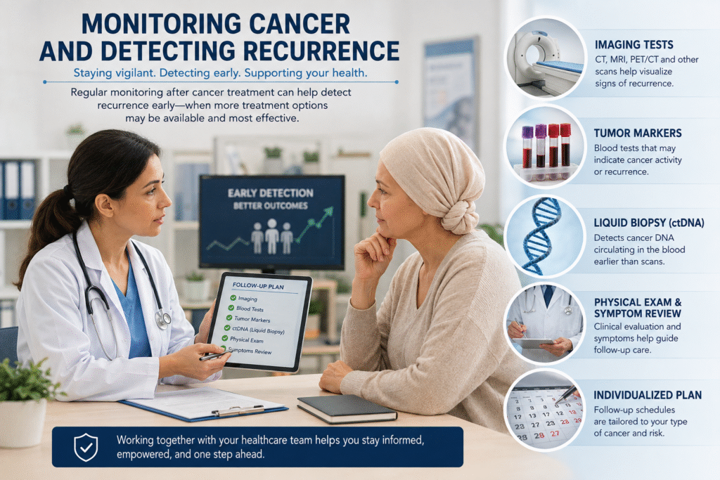
Why Cancer Surveillance Matters
Cancer can recur locally, regionally, or at distant sites.
Local recurrence means that cancer returns in or near the original location. Regional recurrence involves nearby lymph nodes or tissues. Distant recurrence means that malignant cells have established disease elsewhere in the body.
Recurrence occurs because a very small population of malignant cells may sometimes survive initial treatment. These cells may be too few to detect with conventional imaging at the time therapy is completed.
The purpose of surveillance is therefore not simply to order tests. It is to identify clinically meaningful changes at a time when additional treatment may be beneficial.
At the same time, more testing is not automatically better. Surveillance should be based on evidence because excessive imaging and laboratory testing can produce false positive findings, unnecessary biopsies, additional radiation exposure, anxiety, and expense.
Monitoring Cancer and Detecting Recurrence With Imaging
Monitoring cancer and detecting reoccurrence with imaging remains one of the most important tools for following cancer.
Computed tomography, magnetic resonance imaging, positron emission tomography, ultrasound, bone imaging, mammography, and other specialized studies may be used depending upon the cancer.
Imaging can help determine whether a tumor is shrinking, remaining stable, or progressing during treatment. It can also identify new lesions after treatment has been completed. The National Cancer Institute specifically recognizes imaging as a method for assessing treatment effectiveness and monitoring for recurrence.
Different imaging methods answer different questions.
CT provides detailed structural information about many areas of the body.
MRI offers excellent soft tissue detail and is particularly useful in areas such as the brain, spine, liver, breast, pelvis, and prostate.
PET imaging evaluates metabolic or molecular activity and may identify disease that is difficult to distinguish anatomically.
Some cancers also have highly specialized imaging techniques. Prostate specific membrane antigen PET imaging, for example, has changed evaluation of many patients with recurrent or metastatic prostate cancer.
No imaging test detects every malignant cell.
A scan that shows no evidence of disease means that no cancer was detected at the resolution of that test. It does not prove that absolutely no malignant cells remain in the body. This is important to remember when monitoring cancer and detecting reoccurrence.
Blood Tumor Markers
Some cancers release measurable substances into the bloodstream.
These are commonly called tumor markers. Monitoring cancer and detecting recurrence may also include tumor markers when they appropriate.
Examples include prostate specific antigen in prostate cancer, carcinoembryonic antigen in colorectal and certain other cancers, CA 125 in ovarian cancer, CA 19 9 in pancreatic and biliary cancers, and several additional markers used in particular clinical situations.
Serial measurements can sometimes be more useful than a single result.
A rising marker may suggest progression or recurrence. A falling marker during treatment may suggest therapeutic response.
However, tumor markers are imperfect.
Some cancers do not produce them. Some benign conditions can increase them. A normal result does not always exclude cancer, and an elevated result does not automatically establish recurrence.
The National Cancer Institute notes that tumor markers may be measured during therapy to assess response and periodically after treatment to look for recurrence.
Therefore, tumor markers should usually be interpreted together with symptoms, examination findings, imaging, pathology, and the clinical course.
Circulating Tumor DNA and Liquid Biopsy
One of the most important developments in cancer surveillance is circulating tumor DNA.
Tumor cells can release small fragments of their DNA into the bloodstream. This material is known as circulating tumor DNA, or ctDNA.
A blood sample can sometimes detect these tumor derived DNA fragments without requiring another tissue biopsy.
This approach is commonly referred to as liquid biopsy.
Liquid biopsy can potentially provide several types of information.
It may identify molecular alterations in a tumor.
It may help determine whether a targeted treatment is appropriate.
It may allow physicians to observe molecular changes as a cancer evolves.
It may help assess treatment response.
It may sometimes identify evidence of residual or recurrent disease before it becomes obvious on conventional imaging.
The National Cancer Institute has described ctDNA based liquid biopsy as a potentially useful method for monitoring treatment response during and after therapy.
This technology is advancing rapidly, but its clinical role differs among cancers.
Minimal Residual Disease
Minimal residual disease refers to a very small amount of cancer remaining after treatment.
In solid tumors, the closely related term molecular residual disease is increasingly used when tumor derived DNA can be identified despite the absence of visible disease on imaging.
This is an important concept.
A patient may have surgery and subsequently have a CT or PET scan showing no measurable cancer. Yet microscopic malignant cells could still remain.
If ctDNA from that patient’s tumor is detected after treatment, it may suggest a greater probability of recurrence in some cancers.
For example, substantial research is examining postoperative ctDNA in colorectal cancer, breast cancer, lung cancer, and other solid tumors. Current National Cancer Institute trials are evaluating whether ctDNA can identify patients with residual disease and help guide subsequent treatment.
The important limitation is that a negative ctDNA test does not guarantee that no cancer remains.
Tumors vary greatly in how much DNA they release into the bloodstream. Small tumors and tumors in certain anatomical locations may release very little detectable ctDNA.
For this reason, molecular testing does not completely replace imaging or conventional clinical surveillance.
Monitoring Cancer and Detecting Recurrence During Treatment
Surveillance is also important while active treatment is being given.
The oncologist may evaluate several different forms of evidence.
Symptoms may improve or worsen.
Tumor markers may rise or fall.
Imaging may show regression, stability, or progression.
Blood counts and organ function tests may determine whether therapy can safely continue.
Molecular testing may occasionally demonstrate that the cancer is developing new resistance mechanisms.
Increasingly, researchers are studying whether changes in ctDNA during therapy can provide earlier evidence of treatment response than conventional imaging. An active National Cancer Institute trial is evaluating ctDNA monitoring as a way of predicting treatment response in non-small cell lung cancer.
The goal of monitoring and detecting reoccurrence is eventually to identify ineffective treatment sooner and modify therapy before substantial clinical progression occurs.
Why Cancer Can Return After Initially Successful Treatment
Cancer is biologically heterogeneous.
A tumor may contain millions or billions of malignant cells that are not genetically identical.
Treatment may destroy the majority of susceptible cancer cells while allowing a small resistant population to survive.
Those surviving cells can continue evolving.
Over time, they may acquire additional alterations that allow them to grow despite therapies that previously worked.
This is one reason recurrent cancer may behave differently from the original tumor.
When recurrence occurs, physicians may sometimes recommend another tissue biopsy or new molecular testing rather than assuming that the biology remains unchanged.
The estrogen receptor, HER2 status, genomic alterations, resistance mutations, or other characteristics can sometimes change during the evolution of cancer.
Understanding those changes can influence subsequent treatment.
Symptoms Still Matter
Technology does not replace clinical judgment.
New or persistent symptoms can sometimes be the earliest sign that a cancer has returned.
Depending upon the original malignancy, concerning symptoms might include unexplained weight loss, persistent pain, new neurologic symptoms, progressive fatigue, abnormal bleeding, persistent cough, abdominal swelling, changes in bowel habits, new masses, or other unexplained findings.
Most symptoms experienced by cancer survivors are not caused by recurrence.
Nevertheless, important changes should not automatically be dismissed simply because a recent scan was normal.
Cancer surveillance works best when laboratory testing, imaging, symptoms, physical examination, and the patient’s history are considered together.
How Often Should Cancer Be Monitored?
There is no universal schedule.
Some cancers require frequent surveillance during the first several years after treatment because the probability of recurrence is greatest during that period.
Other cancers can recur many years later and therefore require longer follow up.
The National Cancer Institute notes that follow up frequency depends upon cancer type, treatments received, overall health, and treatment related complications. Many patients are initially seen more frequently and then at longer intervals as time passes.
The surveillance schedule should therefore be based on the specific malignancy rather than a generic cancer follow up program.
More Testing Is Not Always Better
One of the difficult parts of cancer surveillance is determining when a test will actually improve outcomes.
Finding something earlier is only valuable if acting on that information benefits the patient.
For example, studies of colorectal cancer surveillance have examined whether more frequent testing improves survival. NCI has reported research showing that substantially increasing the frequency of follow up testing does not necessarily produce better outcomes for every patient.
This principle is important.
A good surveillance program is not the program with the largest number of tests.
It is the program that uses the correct tests at the correct intervals for the individual patient’s cancer.
Monitoring Cancer and Detecting Recurrence Is Becoming More Personalized
The future of cancer surveillance is moving beyond simply scheduling a CT scan every several months.
Modern surveillance increasingly combines pathology, imaging, conventional tumor markers, molecular profiling, and circulating tumor DNA.
The ultimate objective is to recognize recurrence at the earliest clinically meaningful stage while avoiding unnecessary procedures.
This approach may eventually allow surveillance to become increasingly personalized.
A patient with persistent postoperative ctDNA may need a different strategy from someone with repeatedly negative molecular testing.
A patient whose tumor has a specific molecular alteration may benefit from a specialized blood assay.
Another patient may be better monitored through imaging because the cancer releases little detectable DNA into the bloodstream.
The science is moving toward surveillance based on the biology of the individual tumor rather than simply the organ where the cancer began.
For additional patient information, the National Cancer Institute Follow Up Medical Care resource explains how follow up schedules and testing are individualized after cancer treatment.
For additional information about surveillance after cancer treatment, visit the National Cancer Institute Follow-Up Medical Care resource.
Conclusion
Monitoring cancer does not end when treatment ends.
Cancer surveillance may involve clinical examination, laboratory studies, tumor markers, CT, MRI, PET imaging, specialized imaging, tissue biopsy, and increasingly circulating tumor DNA.
Each method provides different information.
Imaging shows disease that is large enough to be visualized.
Tumor markers may provide biochemical evidence of changing disease activity.
Liquid biopsy may identify molecular information released by malignant cells.
Symptoms and physical examination remain important.
No single test provides complete certainty.
The most effective approach to monitoring cancer and detecting recurrence is therefore to combine the appropriate tools according to the type and biology of the cancer, the treatments received, and the patient’s individual risk.
Earlier detection is valuable when it allows physicians and patients to make better treatment decisions.
Educational Disclaimer
This article is provided for educational purposes only. It does not constitute medical advice, diagnosis, treatment, or prescribing. Cancer surveillance must be individualized according to the specific cancer, stage, pathology, molecular characteristics, treatment history, symptoms, overall health, and current clinical guidelines. Patients should discuss surveillance testing and treatment decisions with their treating oncology team. This information does not establish a physician patient relationship.
Dr. Dean Silver
References
- National Cancer Institute. Follow Up Medical Care. NCI Cancer Survivorship Resources. Updated December 2, 2024.
- National Cancer Institute. Cancer Imaging Basics for Diagnosis and Treatment. National Cancer Institute Division of Cancer Treatment and Diagnosis.
- National Cancer Institute. Tumor Markers. NCI Cancer Diagnosis and Staging. Updated December 7, 2023.
- National Cancer Institute. Tumor Marker Tests in Common Use. Updated December 7, 2023.
- National Cancer Institute. Liquid Biopsy: Using Tumor DNA in Blood to Aid Cancer Care. NCI Cancer Currents. November 8, 2017.
- National Cancer Institute. Blood Test May Help Guide Colorectal Cancer Treatment. NCI Cancer Currents. February 28, 2024.
- National Cancer Institute. Circulating Tumor DNA Based Minimal Residual Disease Detection in Early Stage Breast Cancer. Clinical Trial NCI 2025 05431.
- National Cancer Institute. Circulating Tumor DNA Monitoring for Treatment Response in Non Small Cell Lung Cancer. Clinical Trial NCI 2025 04101.
- National Cancer Institute. Postoperative Circulating Tumor DNA Surveillance in Triple Negative Breast Cancer. Clinical Trial NCI 2025 05695.
- Kim H, et al. Clinical Circulating Tumor DNA Testing for Precision Oncology. Precision and Future Medicine. 2023.
- National Cancer Institute. Do Frequent Follow Up Tests Benefit Colorectal Cancer Survivors? NCI Cancer Currents. June 20, 2018.
- National Cancer Institute. Computed Tomography Scans and Cancer. Updated February 8, 2024.