Understanding your pathology report is one of the most important steps after a cancer diagnosis. This report identifies the type of cancer, describes how abnormal the cells appear, evaluates whether surgery removed the tumor completely, examines lymph nodes and other risk features, and often provides biomarkers that influence treatment.
The problem is that pathology reports are written primarily for physicians and pathologists. They contain technical language, abbreviations, measurements, staging codes, and microscopic descriptions that can be difficult to interpret without explanation.
Many patients see these results in an electronic portal before speaking with their oncologist.
This guide explains the major sections so that you can understand what the report is telling you and what questions to ask next.
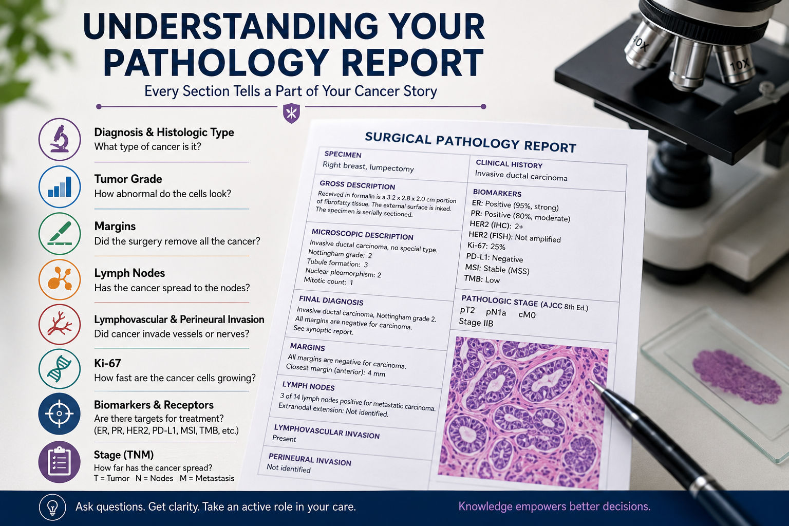
Understanding Your Pathology Report: The Gross Description
The gross description explains what the specimen looked like before it was examined under the microscope.
The pathologist may describe its size, color, shape, texture, orientation, and how the surgical surfaces were inked.
If a tumor is visible, its dimensions are generally recorded in centimeters.
This section is primarily procedural, but tumor size can be important because it may contribute directly to the T component of cancer staging.
The gross description may also identify how close the visible tumor appears to surgical margins before microscopic examination.
The definitive margin assessment, however, comes from examination under the microscope.
Microscopic Description and Final Diagnosis
This is usually the most important section of the pathology report.
The pathologist examines thin sections of tissue under a microscope and determines exactly what type of abnormal cells are present.
Understanding Your Pathology Report and Histologic Type
The histologic type describes what kind of cancer is present based on the tissue and cellular pattern from which it arose.
Examples include adenocarcinoma, squamous cell carcinoma, lymphoma, melanoma, and sarcoma.
An adenocarcinoma arises from gland forming epithelial cells.
Squamous cell carcinoma develops from squamous epithelial cells.
Sarcomas arise from connective tissues such as muscle, bone, fat, or blood vessels.
The histologic type can profoundly influence treatment.
Two tumors located in the same organ may require very different treatment if their histology is different.
In Situ Versus Invasive Cancer
This distinction is fundamental.
In situ means abnormal cells remain confined to the tissue compartment where they originated and have not crossed the basement membrane into surrounding tissue.
Ductal carcinoma in situ of the breast is a familiar example.
In situ disease does not have the same ability to metastasize as invasive cancer because the abnormal cells have not penetrated into surrounding tissue where they can access lymphatic channels and blood vessels.
Invasive cancer means malignant cells have crossed that boundary and entered surrounding tissue.
Once cancer becomes invasive, the biological possibility of lymphatic or blood borne spread exists.
This does not mean metastasis has occurred.
It means the tumor has acquired the anatomical opportunity to spread.
Understanding Your Pathology Report: Tumor Grade
Grade and stage are frequently confused.
They describe two different things.
Grade describes what the cancer cells look like under the microscope.
Stage describes how far the cancer has spread.
A small tumor can be high grade.
A large tumor can be low grade.
Many cancers use a grading system from 1 through 3, although some diseases use different systems.
Grade 1
Grade 1 tumors are usually described as well differentiated.
The cells still resemble the normal tissue from which they originated and generally appear less aggressive microscopically.
Grade 2
Grade 2 means moderately differentiated.
The tumor demonstrates intermediate microscopic features.
Grade 3
Grade 3 tumors are poorly differentiated.
The cells often look very abnormal, have lost many features of their tissue of origin, and may demonstrate increased mitotic activity.
Higher grade usually correlates with more aggressive behavior.
There is an important qualification.
Rapidly dividing tumors may sometimes be more sensitive to chemotherapy because many cytotoxic drugs preferentially affect dividing cells.
Grade therefore contributes important information, but it does not determine treatment by itself.
Specialized Grading Systems
Some cancers use disease specific grading systems.
Breast Cancer and the Nottingham Grade
Breast cancer frequently uses the Nottingham grading system.
It evaluates three features:
Tubule formation.
Nuclear pleomorphism.
Mitotic activity.
Each component receives a score, and the combined score determines the histologic grade.
Prostate Cancer and the Gleason Score
Prostate cancer uses the Gleason grading system and Grade Groups.
The pathologist identifies the most common growth pattern and the second most common pattern.
This is why Gleason 3 plus 4 and 4 plus 3 are not equivalent even though both total 7.
In Gleason 3 plus 4 disease, pattern 3 predominates.
In Gleason 4 plus 3 disease, the more aggressive pattern 4 predominates.
That difference has prognostic and treatment implications.
Understanding Your Pathology Report: Surgical Margins
Margins answer a very practical question.
Did the surgeon remove the entire tumor?
During surgery, the outer surfaces of the specimen are commonly marked with ink. The pathologist then determines whether cancer cells reach the inked edge.
Negative or Clear Margins
Negative margins mean tumor cells are not present at the surgical edge.
You may also see this described as clear margins.
In some settings, an R0 resection refers to complete microscopic removal of the tumor.
Positive Margins
A positive margin means cancer cells extend to the inked surgical surface.
This raises concern that microscopic tumor may remain in the patient.
Depending on the cancer and operation, a positive margin may lead to consideration of additional surgery, radiation therapy, or another treatment strategy.
Close Margins
A close margin means the tumor approaches the edge but does not reach it.
What constitutes an adequate margin differs by cancer type.
For some cancers, a very small negative margin is acceptable.
For others, a larger distance may influence additional treatment.
The actual distance, usually reported in millimeters, can therefore matter.
Understanding Your Pathology Report and Lymph Nodes
The pathology report usually states how many lymph nodes were examined and how many contained cancer.
For example:
0 of 14 lymph nodes positive
or
3 of 18 lymph nodes positive
Lymph node involvement is an important staging and prognostic feature in many solid tumors.
It means cancer cells have reached the regional lymphatic system.
It does not automatically mean distant metastatic disease is present.
Sentinel Lymph Node
The sentinel lymph node is the first lymph node, or group of nodes, expected to receive lymphatic drainage from the tumor.
Sentinel lymph node biopsy allows surgeons to evaluate regional spread while sometimes avoiding removal of a larger number of nodes.
Micrometastases and Isolated Tumor Cells
Very small tumor deposits may be categorized separately.
You may see terms such as isolated tumor cells or micrometastasis.
Their significance depends on the cancer type and clinical setting.
Extranodal Extension
Extranodal extension means cancer has grown through the capsule of an involved lymph node into surrounding tissue.
This can indicate more aggressive regional disease and may influence staging or adjuvant treatment.
Understanding Your Pathology Report: Lymphovascular Invasion
Lymphovascular invasion, or LVI, means cancer cells are visible within small lymphatic channels or blood vessels surrounding the tumor.
This tells us that tumor cells have entered pathways through which cancer can potentially travel.
LVI is generally considered an adverse pathological feature.
However, LVI does not mean that distant metastasis has definitely occurred.
It represents evidence of increased metastatic potential, not proof of metastatic disease.
Perineural Invasion
Perineural invasion, or PNI, means cancer cells are growing along or around nerves.
It is particularly important in cancers including prostate, pancreatic, and head and neck malignancies.
Its prognostic significance depends on the cancer type.
Like lymphovascular invasion, it may influence recommendations for additional treatment.
Understanding Your Pathology Report: Ki-67
Ki-67 is a marker of cell proliferation.
It is expressed in cells that are actively progressing through the cell cycle.
A pathology report may provide a percentage such as:
Ki-67: 5 percent
or
Ki-67: 70 percent
A higher number generally indicates that a larger proportion of tumor cells are actively dividing.
But there is no universal Ki-67 cutoff that applies to every cancer.
Its interpretation is tumor specific.
In neuroendocrine neoplasms, Ki-67 can be central to tumor grading.
In breast cancer, it may provide additional information about proliferation, although laboratory variability has limited the reliability of strict universal thresholds.
A high Ki-67 can indicate a biologically aggressive tumor.
Again, there is an important counterpoint.
Fast dividing tumors may sometimes respond better to chemotherapy than slowly proliferating tumors.
Understanding Your Pathology Report and Biomarkers
Modern pathology reports increasingly contain information that directly identifies possible treatment targets.
Breast cancer provides a clear example.
Estrogen Receptor
ER means estrogen receptor.
ER positive breast cancers may respond to endocrine therapies that reduce estrogen signaling.
Progesterone Receptor
PR means progesterone receptor.
PR provides additional information about hormone receptor biology.
HER2
HER2 is a growth signaling receptor.
HER2 positive breast cancers can respond to HER2 targeted therapies.
HER2 immunohistochemistry is commonly reported as:
0
1+
2+
3+
A score of 3+ generally supports HER2 positivity.
A 2+ result is considered equivocal and typically requires additional testing for gene amplification.
Understanding Your Pathology Report and HER2
HER2 classification has become more nuanced because antibody drug conjugates can benefit some tumors that historically would have been called simply HER2 negative.
The concept of HER2 low generally includes tumors with IHC 1+ or IHC 2+ without HER2 gene amplification.
This matters because drugs such as trastuzumab deruxtecan have demonstrated activity in appropriately selected HER2 low metastatic breast cancers.
This is an example of how pathology terminology can change as new treatments become available.
Understanding Your Pathology Report and Immunotherapy Biomarkers
Some pathology and molecular reports also include markers associated with immunotherapy.
These may include:
PD-L1.
Microsatellite instability.
Mismatch repair proteins.
Tumor mutational burden.
The meaning of these results depends heavily on cancer type, stage, assay, and treatment setting.
For a detailed explanation, see immunotherapy biomarkers:
Understanding Your Pathology Report: TNM Staging
Most solid cancers use some form of the TNM staging system.
T Means Tumor
T describes the size or local extent of the primary tumor.
The categories usually range from T1 to T4, although the exact definitions differ by cancer.
N Means Nodes
N describes regional lymph node involvement.
The categories generally range from N0 through N3.
M Means Metastasis
M describes distant metastatic disease.
M0 means no distant metastasis has been identified within the staging framework.
M1 means distant metastatic disease is present.
The T, N, and M components are then combined with other information to produce an overall stage.
What pT, cT, and ypT Mean
The letters placed before TNM values are important.
c means clinical staging.
This is based on information available before definitive surgery, including physical examination, imaging, endoscopy, biopsy, and other testing.
p means pathological staging.
This is based primarily on examination of tissue removed surgically.
y indicates staging after neoadjuvant treatment.
For example, ypT means the tumor classification was determined pathologically after treatment such as chemotherapy, radiation, or systemic therapy was given before surgery.
These prefixes tell you how the staging information was obtained.
Grade Is Not Stage
This point is worth repeating.
A Grade 3 tumor is not necessarily Stage III cancer.
A Stage I cancer can be Grade 3.
A Stage III cancer can sometimes be relatively low grade.
Grade describes microscopic appearance.
Stage describes the anatomical extent of disease.
They provide different information and should never be used interchangeably.
Common Pathology Terms That Can Sound Frightening
Some pathology language sounds alarming even when it does not necessarily indicate a worse diagnosis.
Atypical
Atypical means cells do not look completely normal.
It does not automatically mean cancer.
Suspicious For
“Suspicious for” means the findings raise concern but are not yet definitive.
Additional stains, tissue, or expert review may be needed.
Necrosis
Necrosis means dead tissue.
Within an untreated tumor, extensive necrosis may sometimes reflect aggressive growth that has outstripped its blood supply.
After treatment, however, tumor necrosis can indicate treatment effect.
The clinical context matters.
Additional Studies Pending
This usually means the pathologist is waiting for immunohistochemical stains, molecular testing, special stains, or another evaluation before issuing the final interpretation.
It is not inherently a bad sign.
Addendum or Amended Report
An addendum may contain additional biomarker, molecular, or immunohistochemistry results obtained after the initial report.
An amended report means something in the original report has been formally changed or clarified.
Read the amendment carefully because it can occasionally alter important diagnostic information.
Understanding Your Pathology Report: Second Opinion Review
Pathology is a highly specialized interpretive field.
In most cases, the diagnosis is straightforward.
But some tumors are difficult to classify.
This is particularly true for rare sarcomas, lymphomas, unusual brain tumors, borderline lesions, and uncommon pathological variants.
A second pathology review at a major cancer center can therefore be reasonable before starting treatment when the diagnosis is rare, unexpected, or uncertain.
The original glass slides or paraffin blocks can usually be sent to another institution for review.
This is routine medical practice.
It is not an insult to the original pathologist.
Questions to Ask About Your Pathology Report
When you meet with your oncologist, ask:
What is the exact histologic diagnosis?
Is the cancer invasive or in situ?
What is the grade?
What is the tumor size?
Are the surgical margins clear?
What is the closest margin?
How many lymph nodes were examined?
How many were positive?
Was lymphovascular invasion present?
Was perineural invasion present?
What is the Ki-67?
Which receptors or biomarkers were tested?
What is the pathological TNM stage?
Do I need additional molecular testing?
Would a second pathology review change anything about my treatment decision?
These questions turn a complicated report into a manageable clinical discussion.
Understanding Your Pathology Report:Frequently Asked Questions
How Long Does a Pathology Report Take?
Routine pathology may be completed within several business days.
The process can take longer if additional immunohistochemical stains, molecular testing, decalcification, specialized consultation, or outside review is needed.
A delay does not necessarily mean that something worse has been found.
What Is the Difference Between Grade and Stage?
Grade describes how abnormal or differentiated the cancer cells appear microscopically.
Stage describes how far the cancer has spread anatomically.
They are separate characteristics.
For detailed standardized cancer staging information, see the National Cancer Institute staging guide.
Can I Get a Second Opinion on My Pathology?
Yes.
Pathology slides and tissue blocks can generally be sent to another pathology department for review.
Second review can be especially valuable in rare tumors, unusual histologies, or cases where the diagnosis will fundamentally determine a major treatment decision.
Does a Positive Margin Mean the Cancer Has Spread?
No.
A positive margin means cancer cells reach the surgical edge of the removed specimen.
It raises concern that microscopic disease may remain locally.
It does not establish distant metastatic disease.
Does Lymphovascular Invasion Mean I Have Metastatic Cancer?
No.
Lymphovascular invasion means cancer cells were observed within small lymphatic or vascular spaces near the tumor.
It indicates an increased biological opportunity for spread but does not prove that distant metastases are present.
Understanding Your Pathology Report:The Bottom Line
Understanding your pathology report means looking at the entire picture rather than focusing on one frightening word or number.
The diagnosis tells you what type of cancer is present.
Grade describes how abnormal and aggressive the cells appear.
Margins tell you whether tumor reaches the edge of the surgical specimen.
Lymph nodes help assess regional spread.
Lymphovascular and perineural invasion provide additional information about tumor behavior.
Ki-67 estimates proliferation.
Receptors and biomarkers may identify treatment targets.
TNM describes the anatomical extent of disease.
No single feature should be interpreted in isolation.
The pathology report becomes most useful when these findings are combined with imaging, molecular testing, your medical history, and the evidence supporting the available treatments.
By Dean R. Silver, MD, MD (H)
Educational Disclaimer
This article is provided for cancer education only. It is not medical advice and does not establish a physician patient relationship. Pathology findings should not be interpreted independently to determine cancer prognosis or to start, stop, or change treatment. The significance of tumor type, grade, margins, lymph nodes, biomarkers, Ki-67, and staging depends on the specific cancer and clinical setting. Review your pathology report with your treating oncologist and, when appropriate, a qualified pathologist.
References
- College of American Pathologists. Cancer Protocol Templates.
- Amin MB, Edge SB, Greene FL, et al., editors. AJCC Cancer Staging Manual. 8th edition. Springer; 2017.
- Rakha EA, Reis Filho JS, Baehner F, et al. Breast cancer prognostic classification in the molecular era: the role of histological grade. Breast Cancer Research. 2010;12(4):207.
- Epstein JI, Egevad L, Amin MB, et al. The 2014 ISUP consensus conference on Gleason grading of prostatic carcinoma. American Journal of Surgical Pathology. 2016;40(2):244 to 252.
- Dowsett M, Nielsen TO, A’Hern R, et al. Assessment of Ki67 in breast cancer: recommendations from the International Ki67 in Breast Cancer Working Group. Journal of the National Cancer Institute. 2011;103(22):1656 to 1664.
- Wolff AC, Somerfield MR, Dowsett M, et al. Human epidermal growth factor receptor 2 testing in breast cancer: ASCO CAP guideline update. Journal of Clinical Oncology. 2023;41(22):3867 to 3872.
- Modi S, Jacot W, Yamashita T, et al. Trastuzumab deruxtecan in previously treated HER2 low advanced breast cancer. New England Journal of Medicine. 2022;387(1):9 to 20.
- Kronz JD, Westra WH, Epstein JI. Mandatory second opinion surgical pathology at a large referral hospital. Cancer. 1999;86(11):2426 to 2435.
- National Cancer Institute. Pathology Reports. National Cancer Institute.