Immunotherapy biomarkers help oncologists estimate whether a cancer may respond to immune checkpoint inhibitors. The three markers’ patients see most often are PD-L1, microsatellite instability, or MSI, and tumor mutational burden, or TMB. They measure different aspects of tumor biology, and none should be interpreted as a simple yes or no test for whether immunotherapy will work.
Checkpoint inhibitors have transformed treatment for many cancers. Some patients experience dramatic and durable responses. Others receive little or no benefit.
The challenge is identifying which tumor is most likely to respond before exposing a patient to treatment.
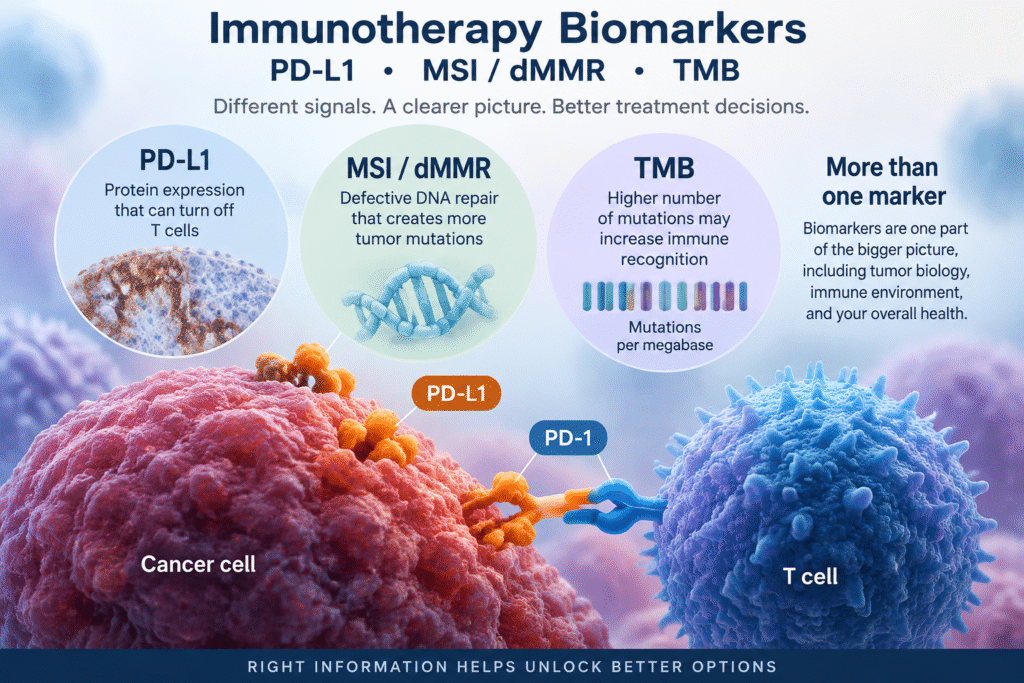
That is the purpose of immunotherapy biomarkers.
How Immunotherapy Biomarkers Relate to Checkpoint Inhibitors
Your immune system contains natural brakes.
T cells express inhibitory receptors including PD-1 and CTLA-4. These checkpoints help prevent excessive immune activation and autoimmune injury.
Cancer cells can exploit this protective system.
Some tumors express PD-L1. When PD-L1 binds to PD-1 on a T cell, an inhibitory signal is transmitted that reduces T cell activity.
The immune cell may recognize the cancer but becomes functionally suppressed.
Checkpoint inhibitors interfere with these inhibitory signals.
Examples include pembrolizumab, or Keytruda; nivolumab, or Opdivo; atezolizumab, or Tecentriq; durvalumab, or Imfinzi; and ipilimumab, or Yervoy.
These drugs generally do not kill cancer cells directly.
They modify immune signaling so that immune cells may resume attacking the tumor.
This distinction helps explain why biomarkers matter.
If there is little useful anti tumor immunity present, simply releasing an immune brake may not be enough.
Immunotherapy Biomarkers and PD-L1
PD-L1 is probably the best known and most confusing immunotherapy biomarker.
PD-L1 is usually measured by immunohistochemistry. A pathologist applies a validated antibody stain to tumor tissue and evaluates how much PD-L1 protein is present and where it is expressed.
The difficulty is that there is no single universal PD-L1 score.
Different cancers use different scoring systems, different assays, and different treatment thresholds.
PD-L1 Immunotherapy Biomarkers: TPS, CPS, and IC
Three scoring systems commonly appear on pathology reports.
Tumor Proportion Score
TPS stands for Tumor Proportion Score.
It measures the percentage of viable tumor cells showing membrane staining for PD-L1.
TPS is commonly used in non small cell lung cancer.
A report might read:
PD-L1 TPS 60 percent
The number must then be interpreted in the context of the specific cancer, treatment regimen, stage, and FDA approved indication. For current assay specific indications and cutoffs, see the FDA companion diagnostic list.
Combined Positive Score
CPS stands for Combined Positive Score.
It incorporates PD-L1 staining in tumor cells and selected tumor associated immune cells.
The basic calculation compares the number of PD-L1 positive tumor and immune cells with the number of viable tumor cells.
CPS is used in several malignancies, including triple negative breast cancer, gastric and gastroesophageal cancers, cervical cancer, and head and neck cancer.
For locally recurrent unresectable or metastatic triple negative breast cancer, pembrolizumab plus chemotherapy has an FDA indication for tumors expressing PD-L1 with CPS 10 or greater, using an FDA approved test.
Immune Cell Score
Some assays evaluate PD-L1 positive immune cells within the tumor microenvironment.
This illustrates why simply telling a patient, “Your PD-L1 is 10,” is inadequate.
You need to know which scoring method produced the number.
Why PD-L1 Is an Imperfect Biomarker
PD-L1 provides useful information, but it does not perfectly predict treatment response.
Some tumors with high PD-L1 expression fail to respond.
Some tumors with low or undetectable PD-L1 expression respond to checkpoint inhibition.
PD-L1 can also vary between a primary tumor and a metastasis.
It may change after chemotherapy or radiation.
Different validated antibody assays can also produce somewhat different results.
For this reason, PD-L1 should be interpreted as one piece of evidence rather than a verdict.
Immunotherapy Biomarkers in Triple Negative Breast Cancer
Triple negative breast cancer provides one of the clearest examples of why the clinical setting matters as much as the biomarker.
In high risk early stage triple negative breast cancer, pembrolizumab can be used with neoadjuvant chemotherapy and continued as adjuvant treatment without requiring PD-L1 positivity.
The KEYNOTE-522 treatment strategy was not restricted to PD-L1 positive disease.
In contrast, for locally recurrent unresectable or metastatic triple negative breast cancer, pembrolizumab plus chemotherapy uses PD-L1 CPS of 10 or greater as a companion diagnostic criterion.
This can appear contradictory.
It is not.
The early stage and metastatic trials addressed different clinical settings and used different eligibility and treatment strategies.
This is why you cannot interpret PD-L1 without knowing the stage and treatment setting.
MSI and Mismatch Repair Deficiency
Among immunotherapy biomarkers, microsatellite instability and mismatch repair deficiency have particularly strong biological rationale.
Cells constantly copy DNA.
Copying produces mistakes.
The mismatch repair system acts as a proofreading mechanism that identifies and repairs many of these errors.
Four important mismatch repair proteins are:
MLH1
MSH2
MSH6
PMS2
When this repair system fails, mutations accumulate.
Repetitive DNA sequences called microsatellites become especially unstable.
A tumor demonstrating this pattern is called microsatellite instability high, or MSI-H.
When the repair proteins themselves are absent or dysfunctional, the tumor is described as mismatch repair deficient, or dMMR.
Immunotherapy Biomarkers and Why MSI-H Matters
MSI-H and dMMR tumors accumulate large numbers of genetic abnormalities.
Those abnormalities can produce abnormal proteins called neoantigens.
The immune system may recognize these proteins as foreign.
This can make the cancer unusually visible to T cells.
Checkpoint inhibition can therefore be particularly effective in some MSI-H or dMMR tumors because the immune system already has many abnormal targets to recognize.
Pembrolizumab’s development in MSI-H and dMMR cancers helped establish the concept of treating cancers according to a molecular characteristic rather than only according to the organ where the cancer began. FDA companion diagnostic approvals now include testing for MSI-H status in solid tumors that may benefit from pembrolizumab.
How MSI and dMMR Are Tested
Mismatch repair status can be assessed in several ways.
Immunohistochemistry examines whether the MLH1, MSH2, MSH6, and PMS2 proteins are present in tumor tissue.
PCR based testing can directly evaluate microsatellite instability.
Next generation sequencing can also identify MSI and associated genomic abnormalities.
Abnormal mismatch repair testing can also raise the possibility of Lynch syndrome.
That is important because Lynch syndrome is inherited and can affect cancer risk for the patient and biological relatives.
Tumor testing suggesting Lynch syndrome may therefore lead to germline genetic evaluation.
Tumor Mutational Burden
Tumor mutational burden, or TMB, asks a different question.
It estimates the number of somatic mutations present within a defined amount of tumor DNA.
TMB is usually expressed as:
mutations per megabase
The theory is straightforward.
The more mutations a tumor carries, the greater the potential number of abnormal proteins or neoantigens it may produce.
More neoantigens may increase the probability that immune cells recognize the cancer.
Immunotherapy Biomarkers and TMB High
For the FDA tumor agnostic pembrolizumab indication, TMB high is defined as at least 10 mutations per megabase, determined by an FDA approved test, for unresectable or metastatic solid tumors that have progressed after prior treatment and have no satisfactory alternative treatment options.
This number requires some perspective.
Ten mutations per megabase is a regulatory threshold.
It is not a biological cliff where a tumor at 9 mutations per megabase is completely different from a tumor at 10.
Different cancers also have different baseline mutation rates.
The predictive meaning of TMB therefore varies by tumor type.
Why TMB Numbers Can Differ Between Laboratories
TMB is more complicated than simply counting mutations.
Different sequencing panels analyze different amounts of DNA.
Laboratories may use different methods for filtering germline variants and sequencing artifacts.
Bioinformatic calculations also differ.
Therefore, a TMB value from one sequencing platform may not be perfectly interchangeable with a value generated by another platform.
The platform matters.
The cancer type matters.
And the clinical indication matters.
Tissue TMB Versus Blood TMB
TMB can sometimes be estimated from circulating tumor DNA.
This is often called blood TMB.
It can be useful when adequate tumor tissue is unavailable.
But blood based measurements introduce additional variables.
A tumor must shed enough DNA into circulation to generate a reliable measurement.
Low circulating tumor DNA can reduce confidence in the result.
For this reason, tissue based TMB remains more established for many clinical applications.
Why Immunotherapy Biomarkers Can Still Be Wrong
Even when PD-L1, MSI, and TMB have all been measured, immunotherapy response remains difficult to predict.
Cancer immunity involves far more than three laboratory values.
Antigen Presentation Can Fail
T cells must first be able to recognize the tumor.
Cancer cells present antigens through HLA molecules.
Genes such as B2M participate in this process.
If antigen presentation machinery is lost, the immune system may have difficulty recognizing the tumor.
A checkpoint inhibitor cannot release a meaningful immune response if the T cell cannot adequately see its target.
JAK Signaling Can Fail
JAK1 and JAK2 participate in interferon signaling.
Alterations in this pathway have been described as mechanisms of resistance to PD-1 blockade.
A tumor can therefore possess apparently favorable biomarkers while harboring another alteration that weakens the immune response.
Some Tumors Are Immunologically Cold
Some tumors contain very few tumor infiltrating T cells.
These are sometimes called immunologically cold tumors.
If there are few active T cells present, removing an inhibitory checkpoint may produce little effect.
The Tumor Microenvironment Matters
Cancer cells live inside an ecosystem.
Suppressive immune cells, abnormal blood vessels, hypoxia, metabolic competition, and tumor acidity can all influence T cell function.
This means the same PD-L1 value may have different significance in different tumor microenvironments.
The Gut Microbiome May Matter
The intestinal microbiome has also been associated with response to checkpoint inhibitor treatment.
Studies have found associations between antibiotic exposure near immunotherapy initiation and poorer outcomes in several cancers.
This remains an active field of investigation and should not be interpreted as a reason to avoid medically necessary antibiotics.
Why Several Immunotherapy Biomarkers Are Better Than One
The future of immunotherapy prediction is unlikely to depend on one marker.
PD-L1 tells us something about checkpoint signaling.
MSI and mismatch repair tell us about DNA repair failure.
TMB provides information about mutation burden.
Antigen presentation genes tell us whether the immune system can see the cancer.
The tumor microenvironment tells us whether T cells can function once they arrive.
The microbiome may influence systemic immune activity.
These systems interact.
For that reason, modern molecular oncology increasingly examines composite patterns rather than interpreting a single laboratory number in isolation.
For a broader discussion of how these findings fit into treatment decisions, see cancer treatment approaches.
Questions to Ask About Your Immunotherapy Biomarkers
When reviewing a molecular or pathology report, useful questions include:
Which PD-L1 scoring system was used?
Was the result TPS, CPS, or another score?
Which PD-L1 antibody assay was performed?
Was MSI tested?
Was mismatch repair tested by immunohistochemistry?
Was TMB reported?
What sequencing platform produced the TMB result?
Was the specimen from the original tumor or a newer metastasis?
How old is the tissue?
Does PD-L1 actually determine eligibility for immunotherapy in my cancer and stage?
These questions can be more useful than simply asking whether a biomarker is positive or negative.
Frequently Asked Questions
Can Immunotherapy Work if PD-L1 Is Negative?
Yes, in some settings.
PD-L1 is not required for every checkpoint inhibitor indication.
For example, pembrolizumab treatment in high risk early stage triple negative breast cancer is not restricted to PD-L1 positive tumors.
MSI-H, dMMR, TMB high status, cancer type, stage, treatment combination, and other factors may also influence eligibility.
Which Immunotherapy Biomarkers Can Be Tested From Blood?
MSI and TMB can sometimes be evaluated through circulating tumor DNA.
However, tissue based testing remains more established in many situations.
PD-L1 is fundamentally a protein expression test evaluated in tissue by immunohistochemistry.
Can PD-L1 Change Over Time?
Yes.
PD-L1 expression can differ between the primary cancer and metastatic sites and may change after treatment.
If an important treatment decision depends heavily on PD-L1, the age and location of the specimen may therefore matter.
Is TMB 10 Automatically a Reason to Use Immunotherapy?
No.
The FDA TMB-high indication has specific clinical requirements, including disease setting, previous treatment, lack of satisfactory alternatives, and use of an FDA approved test.
A laboratory number should always be interpreted within the full clinical context.
The Bottom Line
Immunotherapy biomarkers are valuable, but none is perfect.
PD-L1 measures one aspect of immune checkpoint biology.
MSI and dMMR identify defective DNA repair that can make a tumor highly immunogenic.
TMB estimates the number of mutations that may generate abnormal proteins recognizable by the immune system.
But cancer immunity is more complex than any one laboratory measurement.
The strongest interpretation combines the biomarker results with the cancer type, stage, prior treatment, molecular profile, tumor microenvironment, and evidence supporting the specific checkpoint inhibitor being considered.
The goal is not simply to ask whether PD-L1, MSI, or TMB is positive.
The better question is:
What does this biomarker mean for this particular cancer, at this particular stage, with this particular treatment?
Dr. Dean R. Silver, MD, MD(H)
Educational Disclaimer
This article is provided for cancer education only. It is not medical advice and does not establish a physician patient relationship. Immunotherapy biomarkers such as PD-L1, MSI, dMMR, and TMB should not be used independently to start, stop, or change cancer treatment. These results must be interpreted together with the cancer type, stage, pathology, prior treatments, overall molecular profile, and current clinical guidelines. Treatment decisions should be discussed with your treating oncologist.
References
- U.S. Food and Drug Administration. List of FDA Authorized Companion Diagnostic Devices. Updated 2026.
- Schmid P, Cortes J, Dent R, et al. Event free survival with pembrolizumab in early triple negative breast cancer. KEYNOTE-522. New England Journal of Medicine. 2022;386(6):556 to 567.
- Cortes J, Rugo HS, Cescon DW, et al. Pembrolizumab plus chemotherapy in advanced triple negative breast cancer. KEYNOTE-355. New England Journal of Medicine. 2022;387(3):217 to 226.
- Reck M, Rodríguez Abreu D, Robinson AG, et al. Pembrolizumab versus chemotherapy for PD-L1 positive non small cell lung cancer. New England Journal of Medicine. 2016;375(19):1823 to 1833.
- Le DT, Durham JN, Smith KN, et al. Mismatch repair deficiency predicts response of solid tumors to PD-1 blockade. Science. 2017;357(6349):409 to 413.
- Marabelle A, Le DT, Ascierto PA, et al. Efficacy of pembrolizumab in patients with noncolorectal high microsatellite instability or mismatch repair deficient cancer. Journal of Clinical Oncology. 2020;38(1):1 to 10.
- Marabelle A, Fakih M, Lopez J, et al. Association of tumour mutational burden with outcomes in patients with advanced solid tumours treated with pembrolizumab. Lancet Oncology. 2020;21(10):1353 to 1365.
- Zaretsky JM, Garcia Diaz A, Shin DS, et al. Mutations associated with acquired resistance to PD-1 blockade in melanoma. New England Journal of Medicine. 2016;375(9):819 to 829.
- Hirsch FR, McElhinny A, Stanforth D, et al. PD-L1 immunohistochemistry assays for lung cancer: results from Phase 1 of the Blueprint PD-L1 IHC Assay Comparison Project. Journal of Thoracic Oncology. 2017;12(2):208 to 222.
- Routy B, Le Chatelier E, Derosa L, et al. Gut microbiome influences efficacy of PD-1 based immunotherapy against epithelial tumors. Science. 2018;359(6371):91 to 97.